Welcome to our article on the 10 week scan and its ability to determine the gender of a baby. Many expectant parents are eager to know the sex of their baby as early as possible, and the 10 week scan is often one of the first opportunities to find out.
In this article, we’ll explore the accuracy of gender determination at 10 weeks, the technology behind the scan, and what to expect during the procedure. Whether you’re curious about the gender of your baby or simply interested in learning more about the 10 week scan, read on to discover everything you need to know.
Can You Tell Gender At 10 Week Scan?
At 10 weeks, it is possible to determine the gender of a fetus using ultrasound, although it may not always be accurate. The genitalia of a male fetus and a female fetus start to develop around the same time, but they look very similar and are often difficult to distinguish until later in the pregnancy.
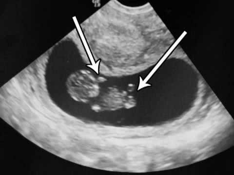
During a 10-week ultrasound scan, the sonographer will look for the presence of a genital tubercle, which is a small protrusion between the legs of the fetus that will eventually develop into either a penis or a clitoris. The angle of the genital tubercle in relation to the spine can also be an indicator of the gender of the fetus. A genital tubercle angled upwards towards the head is more likely to be a male fetus, while an angle that points towards the feet is more likely to indicate a female fetus.
However, it is important to note that the accuracy of determining gender at 10 weeks is lower compared to later in the pregnancy. The fetal genitalia can be difficult to see clearly, and the angle of the genital tubercle may be difficult to determine accurately. Therefore, it is possible to have a wrong or inconclusive gender determination at 10 weeks. In general, it is more common to determine the gender of a fetus with greater accuracy during a routine ultrasound around 18-20 weeks of pregnancy.
How Is Gender Determined During The 10-Week Scan?
During the 10-week scan, also known as the dating scan, a sonographer will use an ultrasound machine to take images of the developing fetus. Gender determination at this stage is possible through a process called “nub theory”. This theory is based on the appearance of the genital tubercle, which is the early fetal structure that eventually develops into either a penis or clitoris.
The genital tubercle is visible on ultrasound images around the 11th week of pregnancy. At this stage, it appears as a small protrusion between the legs of the fetus. The nub theory involves measuring the angle between the genital tubercle and the spine. A genital tubercle that is angled upwards at 30 degrees or more is more likely to indicate a male fetus, while an angle of less than 30 degrees is more likely to indicate a female fetus.
It’s important to note that the accuracy of gender determination at the 10-week scan can vary, and it’s not always possible to get a clear view of the genital tubercle. In some cases, the sonographer may not be able to determine the gender at all. It’s also worth remembering that the primary purpose of the 10-week scan is to check the overall health and development of the fetus, not to determine the gender. If you’re hoping to find out the gender of your baby, it’s best to discuss your options with your healthcare provider.
Ultrasound Technology And Its Capabilities Differences In Genital Development Between Male And Female Fetuses
Ultrasound technology has revolutionized prenatal care, allowing healthcare providers to closely monitor the development of a fetus throughout pregnancy. One of the most exciting capabilities of ultrasound is the ability to determine the gender of a fetus, which is typically possible by around 16-20 weeks of pregnancy.
Differences in genital development between male and female fetuses can be seen on ultrasound images. In a male fetus, the genital tubercle typically begins to elongate and form a penis around the 9th week of pregnancy. By the 14th week, the penis is usually clearly visible on ultrasound. In a female fetus, the genital tubercle remains shorter and eventually forms the clitoris.
During an ultrasound, the sonographer will look for these differences in genital development to determine the gender of the fetus. However, it’s important to remember that the accuracy of gender determination can vary, especially in the early weeks of pregnancy. Factors such as the position of the fetus and the skill of the sonographer can impact the ability to determine the gender.
Despite the potential for variation, ultrasound technology has made it easier than ever to monitor fetal development and detect any potential issues. By providing a detailed view of the fetus, ultrasound allows healthcare providers to identify any abnormalities and develop a treatment plan if necessary. Overall, ultrasound technology plays a vital role in ensuring the health and well-being of both mother and baby during pregnancy.
How A Sonographer Determines The Gender?
Determining the gender of a fetus during an ultrasound scan is an exciting moment for many expectant parents. But how does a sonographer go about determining the gender?
Typically, gender determination is based on the appearance of the external genitalia, which can be seen on ultrasound images. During the ultrasound scan, the sonographer will examine the fetus in detail, looking for differences in the genital area between male and female fetuses.
In a male fetus, the genital tubercle will begin to elongate and form a penis around the 9th week of pregnancy. By the 14th week, the penis is usually clearly visible on ultrasound. In contrast, the genital tubercle in a female fetus will remain shorter and eventually form the clitoris.
The sonographer will look for these differences in genital development to determine the gender of the fetus. However, it’s important to remember that the accuracy of gender determination can vary, especially in the early weeks of pregnancy. Factors such as the position of the fetus and the skill of the sonographer can impact the ability to determine the gender.
It’s also important to note that the primary purpose of the ultrasound scan is to check the overall health and development of the fetus, not to determine the gender. While gender determination can be an exciting part of the scan, it’s not always possible to get a clear view of the genital area, and not all parents want to know the gender in advance.
In summary, a sonographer determines the gender of a fetus by examining the genital area for differences between male and female development. While it’s not always possible to determine the gender with 100% accuracy, ultrasound technology has made it easier than ever to monitor fetal development and ensure the health and well-being of both mother and baby.
Accuracy Of Determining Gender At The 10-Week Scan
Determining the gender of a fetus during the 10-week scan can be an exciting moment for expectant parents. However, it’s important to understand that the accuracy of gender determination at this stage can vary.
The 10-week scan is typically used to confirm the due date and assess the overall health and development of the fetus. While it is possible to determine the gender at this stage, the genital tubercle is still developing, and the fetus may not yet have fully-formed genitals.
The accuracy of gender determination during the 10-week scan is based on the “nub theory”, which involves measuring the angle between the genital tubercle and the spine. While a genital tubercle that is angled upwards at 30 degrees or more is more likely to indicate a male fetus, and an angle of less than 30 degrees is more likely to indicate a female fetus, the accuracy of this method can be affected by a number of factors.
Factors that can impact the accuracy of gender determination include the position of the fetus, the quality of the ultrasound image, and the skill of the sonographer. Additionally, not all fetuses develop at the same rate, which means that the appearance of the genital tubercle can vary from one fetus to another.
Overall, while gender determination at the 10-week scan can be an exciting moment, it’s important to keep in mind that the primary purpose of the scan is to assess the health and development of the fetus. If you’re hoping to find out the gender of your baby, it’s best to discuss your options with your healthcare provider and consider waiting until a later stage of pregnancy when gender determination is likely to be more accurate.
Factors That Can Affect Accuracy
Determining the gender of a fetus during pregnancy is an exciting moment for expectant parents. However, it’s important to understand that the accuracy of gender determination can be affected by a number of factors.
One factor that can impact the accuracy of gender determination is the position of the fetus. If the fetus is not in the right position, it may be difficult to get a clear view of the genital area. In some cases, the fetus may be in a position where the genital area is obscured, making it impossible to determine the gender.
Another factor that can affect accuracy is the quality of the ultrasound image. If the image is blurry or unclear, it may be difficult to see the genital area and make an accurate determination. In some cases, a repeat scan may be necessary to obtain a better image.
The skill and experience of the sonographer can also impact the accuracy of gender determination. A sonographer who is less experienced may have difficulty identifying the genital area or interpreting what they see. It’s important to choose a qualified and experienced sonographer to perform the scan.
Finally, it’s important to note that not all fetuses develop at the same rate. This means that the appearance of the genital area can vary from one fetus to another, and it may not always be possible to make an accurate determination of the gender.
In summary, while ultrasound technology has made it easier than ever to monitor fetal development and determine the gender of a fetus, it’s important to understand that accuracy can be affected by a number of factors. If you’re hoping to find out the gender of your baby, it’s best to discuss your options with your healthcare provider and consider waiting until a later stage of pregnancy when gender determination is likely to be more accurate.
Studies And Statistics On The Accuracy Of The 10-Week Scan For Gender Determination
There have been several studies conducted on the accuracy of gender determination during the 10-week scan, and the results have been mixed.
One study published in the Journal of Ultrasound in Medicine found that the accuracy of gender determination using the “nub theory” was around 70 percent at 11 weeks gestation, and increased to around 98 percent at 14 weeks gestation. However, another study published in the same journal found that the accuracy of gender determination at 11-13 weeks gestation was only around 57 percent.
A larger study published in Ultrasound in Obstetrics and Gynecology found that the accuracy of gender determination using the “nub theory” was around 75 percent at 11-12 weeks gestation, and increased to around 96 percent at 13-14 weeks gestation.
Overall, while the accuracy of gender determination during the 10-week scan may not be as high as during later stages of pregnancy, it’s important to keep in mind that the primary purpose of the scan is to assess the health and development of the fetus. If you’re hoping to find out the gender of your baby, it’s best to discuss your options with your healthcare provider and consider waiting until a later stage of pregnancy when gender determination is likely to be more accurate
The Importance Of A Follow-Up Scan
A follow-up scan is an important step in monitoring the health and development of a fetus, particularly if the initial scan raises any concerns or if there is a need for further evaluation. This is especially true for gender determination during the 10-week scan, as accuracy can be impacted by a number of factors.
A follow-up scan may be recommended if the initial scan was unclear or if the fetus was in a difficult position that made it difficult to determine the gender. Additionally, a follow-up scan may be recommended if there are any concerns about the health or development of the fetus, such as a potential abnormality or growth restriction.
It’s important to note that a follow-up scan may not always be necessary, and the decision to schedule one will depend on the individual circumstances of the pregnancy. If a follow-up scan is recommended, it may be scheduled for a later stage of pregnancy when gender determination is likely to be more accurate.
In summary, a follow-up scan is an important step in monitoring the health and development of a fetus, particularly if there are any concerns or if accuracy of gender determination during the 10-week scan is in question. If you’re unsure whether a follow-up scan is necessary or have any concerns about the health of your pregnancy, it’s best to discuss your options with your healthcare provider.
The Limitations And Risks Of Determining Gender During The 10-Week Scan
While ultrasound technology has made it possible to determine the gender of a fetus as early as the 10-week scan, there are limitations and risks associated with this procedure.
One limitation is the accuracy of gender determination, which can be impacted by factors such as the position of the fetus and the quality of the ultrasound image. In some cases, the gender may not be visible or may be misinterpreted due to fetal development.
Another limitation is the risk of unnecessary anxiety or disappointment. If a parent becomes attached to the idea of a particular gender and then finds out later in pregnancy that the initial determination was incorrect, it can lead to disappointment and emotional distress.
There are also risks associated with any medical procedure, including the 10-week scan. While ultrasound is generally considered safe, there is a small risk of harm to the fetus if it is performed for extended periods or with high-frequency sound waves.
It’s important to keep in mind that while gender determination can be an exciting moment for expectant parents, it is not a necessary part of prenatal care. The primary purpose of the 10-week scan is to assess the health and development of the fetus, and any information about gender should be considered a secondary benefit.
If you’re considering a 10-week scan for gender determination, it’s important to discuss the risks and limitations with your healthcare provider and carefully consider whether it’s the right choice for you and your pregnancy.
The Potential For Misidentification
There is a potential for misidentification when determining the gender of a fetus during the 10-week scan. This can occur due to a number of factors, including the position of the fetus, the quality of the ultrasound image, and the interpretation of the sonographer.
One common issue is that the fetal genitalia may not yet be fully developed at 10 weeks gestation, which can make gender determination more challenging. Additionally, the position of the fetus can impact visibility and make it more difficult to determine the gender with certainty.
It’s important to keep in mind that even experienced sonographers can misidentify the gender of a fetus during the 10-week scan. In some cases, the sonographer may misinterpret the image and provide incorrect information about the gender.
To reduce the risk of misidentification, it’s important to choose a qualified and experienced sonographer who is familiar with the limitations and challenges of gender determination during the 10-week scan. Additionally, it may be helpful to consider a follow-up scan at a later stage of pregnancy when gender determination is likely to be more accurate.
Overall, while misidentification is a potential risk when determining the gender of a fetus during the 10-week scan, it’s important to keep in mind that the primary purpose of the scan is to assess the health and development of the fetus. If you’re considering a 10-week scan for gender determination, it’s important to discuss the potential risks with your healthcare provider and make an informed decision based on your individual circumstances.
The Potential For Misinterpretation
Misinterpretation is another potential issue that can arise when determining the gender of a fetus during the 10-week scan. This can occur due to a variety of factors, including the quality of the ultrasound image, the position of the fetus, and the interpretation of the sonographer.
One common issue is that the images produced during the 10-week scan can be more difficult to interpret due to the size and position of the fetus. This can make it more challenging to distinguish between male and female genitalia with certainty.
Additionally, the interpretation of the sonographer can also impact the accuracy of gender determination. Even experienced sonographers can make mistakes or misinterpret images, which can lead to incorrect information about the gender.
To reduce the risk of misinterpretation, it’s important to choose a qualified and experienced sonographer who is familiar with the nuances of gender determination during the 10-week scan. Additionally, it may be helpful to consider a follow-up scan at a later stage of pregnancy when gender determination is likely to be more accurate.
It’s important to keep in mind that while misinterpretation is a potential risk, the primary purpose of the 10-week scan is to assess the health and development of the fetus. If you’re considering a 10-week scan for gender determination, it’s important to discuss the potential risks with your healthcare provider and make an informed decision based on your individual circumstances.
The Ethical Implications Of Determining Gender Early In Pregnancy
Determining the gender of a fetus early in pregnancy can raise ethical concerns and implications. One potential concern is the use of gender determination for non-medical reasons, such as gender selection or gender preference.
In some cultures, there is a strong preference for a particular gender, and gender determination early in pregnancy may lead to selective abortion or other forms of discrimination. This can have serious ethical implications and contribute to gender inequality.
Additionally, there is a potential for gender stereotypes and expectations to be reinforced through early gender determination. This can impact the way parents interact with their child and can have long-term effects on the child’s development and self-esteem.
It’s important to approach gender determination during the 10-week scan with caution and consideration of the potential ethical implications. It’s also important to remember that gender is a complex and multifaceted aspect of identity that extends beyond biological sex, and that the sex assigned at birth does not necessarily determine a person’s gender identity or expression.
If you have concerns or questions about the ethical implications of early gender determination, it’s important to discuss them with your healthcare provider and seek out resources and support to make informed decisions about your pregnancy and parenting.
Alternatives To The 10-Week Scan For Gender Determination
There are several alternatives to the 10-week scan for gender determination, which may be more accurate and reliable. These alternatives include:
- The 14-week scan: The 14-week scan, also known as the mid-pregnancy or anatomy scan, is a more comprehensive ultrasound scan that assesses the development of the fetus and can also be used to determine the gender. At this stage, the genitalia are more fully developed, making gender determination more accurate.
- Non-invasive prenatal testing (NIPT): NIPT is a blood test that can detect fetal DNA in the mother’s blood and is used to screen for certain genetic conditions. NIPT can also be used to determine the sex of the fetus with a high degree of accuracy.
- Amniocentesis: Amniocentesis is a diagnostic test that involves taking a sample of the amniotic fluid surrounding the fetus. This test can be used to diagnose certain genetic conditions and can also be used to determine the gender of the fetus with a high degree of accuracy.
It’s important to keep in mind that these alternatives may not be suitable or necessary for all pregnancies, and may carry their own risks and limitations. It’s important to discuss the options with your healthcare provider and make an informed decision based on your individual circumstances and preferences.
Ultimately, the decision to determine the gender of your fetus is a personal one and should be made with careful consideration of the potential risks, benefits, and ethical implications.
Non-Invasive Prenatal Testing (Nipt)
Non-Invasive Prenatal Testing (NIPT) is a blood test that can be performed during pregnancy to screen for certain genetic conditions in the fetus. NIPT works by analyzing the fetal DNA that is present in the mother’s blood.
NIPT is considered a non-invasive test because it does not involve any risk to the fetus. Unlike other prenatal tests, such as amniocentesis or chorionic villus sampling, NIPT does not require the insertion of a needle into the uterus.
One of the most common uses of NIPT is to screen for chromosomal abnormalities, such as Down syndrome, trisomy 18, and trisomy 13. NIPT can also be used to determine the sex of the fetus with a high degree of accuracy, making it a useful alternative to the 10-week scan for gender determination.
NIPT is generally considered to be very accurate, with a low rate of false positives and false negatives. However, it’s important to keep in mind that NIPT is a screening test, and a positive result does not necessarily mean that the fetus has a genetic condition.
If NIPT results are positive, further diagnostic testing, such as amniocentesis or chorionic villus sampling, may be recommended to confirm the diagnosis.
It’s important to discuss the options for prenatal testing, including NIPT, with your healthcare provider and make an informed decision based on your individual circumstances and preferences.
Chorionic Villus Sampling (CVS)
Chorionic villus sampling (CVS) is a prenatal diagnostic test that can be performed to detect certain genetic conditions in a developing fetus. The test involves removing a small sample of tissue from the placenta, which contains genetic material from the fetus.
CVS is typically performed between 10-13 weeks of pregnancy and can be used to detect chromosomal abnormalities, such as Down syndrome, as well as certain genetic disorders, such as cystic fibrosis and sickle cell disease.
CVS is considered a diagnostic test, meaning it can provide a definitive diagnosis of a genetic condition. However, it is an invasive procedure that carries some risk to both the fetus and the mother. Possible risks include miscarriage, infection, and damage to the fetus.
CVS is typically only recommended for women who have an increased risk of having a child with a genetic condition. This may include women who are over the age of 35, have a family history of genetic disorders, or have had abnormal results from other prenatal tests.
It’s important to discuss the risks and benefits of CVS with your healthcare provider before deciding whether to have the test. In some cases, non-invasive tests such as NIPT may be recommended as a first-line screening test before considering more invasive diagnostic tests such as CVS.
Amniocentesis
Amniocentesis is a prenatal diagnostic test that involves removing a small sample of amniotic fluid from the uterus to test for certain genetic conditions in the developing fetus. The test is typically performed between 15-20 weeks of pregnancy.
During the procedure, a thin needle is inserted through the mother’s abdomen and into the amniotic sac. A small amount of fluid is then removed and sent to a laboratory for analysis.
Amniocentesis can be used to detect chromosomal abnormalities, such as Down syndrome, as well as certain genetic disorders, such as cystic fibrosis and sickle cell disease. The test is considered a diagnostic test, meaning it can provide a definitive diagnosis of a genetic condition.
However, amniocentesis is an invasive procedure that carries some risk to both the fetus and the mother. Possible risks include miscarriage, infection, and injury to the fetus.
Amniocentesis is typically only recommended for women who have an increased risk of having a child with a genetic condition. This may include women who are over the age of 35, have a family history of genetic disorders, or have had abnormal results from other prenatal tests.
It’s important to discuss the risks and benefits of amniocentesis with your healthcare provider before deciding whether to have the test. In some cases, non-invasive tests such as NIPT may be recommended as a first-line screening test before considering more invasive diagnostic tests such as amniocentesis.
Conclusion
In conclusion, while it’s possible to determine the gender of a baby at the 10 week scan, the accuracy can vary depending on a number of factors. It’s important to keep in mind that the main purpose of this early ultrasound is to assess the health and development of the baby, rather than to determine the sex. However, if you’re eager to know the gender of your baby, your doctor or ultrasound technician may be able to provide some insights. As always, it’s best to discuss any questions or concerns with your healthcare provider to ensure the best possible outcome for you and your baby. Thank you for reading our article on the 10 week scan and gender determination. We hope you found it informative and helpful!